☎ Tick-related emergency?   Poison Control: 1-800-222-1222  — 24/7 Confidential
Species ID Pathogens Clinical Data Treatment Alpha-gal Emerging Viruses Pitfalls Post-Bite Reporting
Section 1

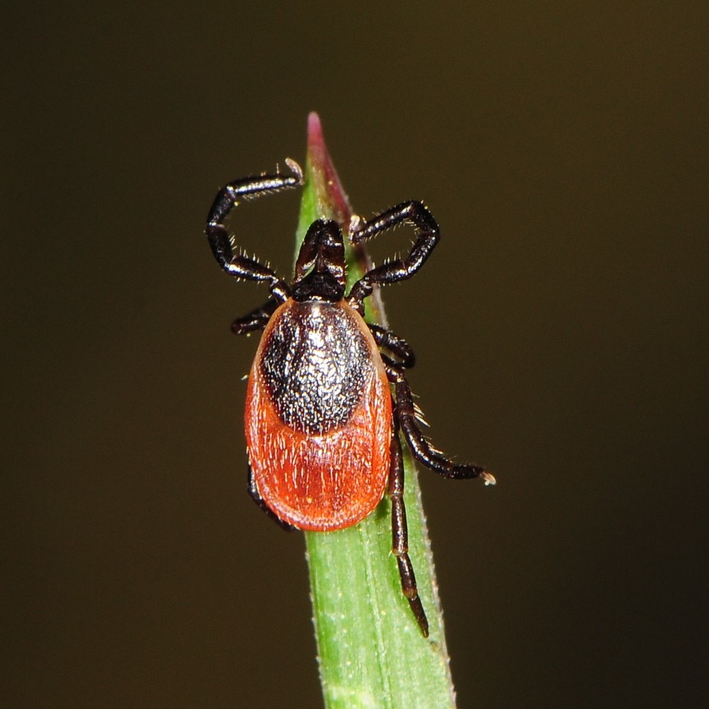
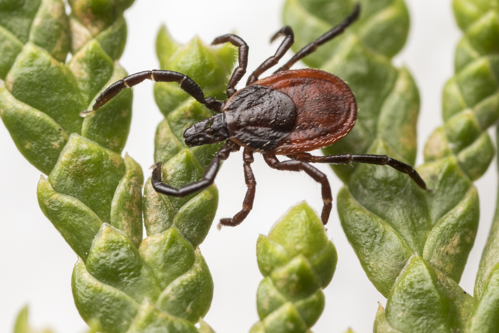
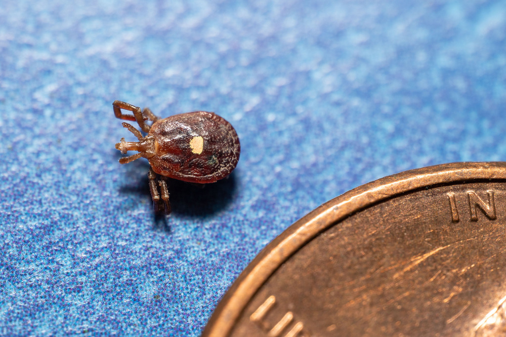
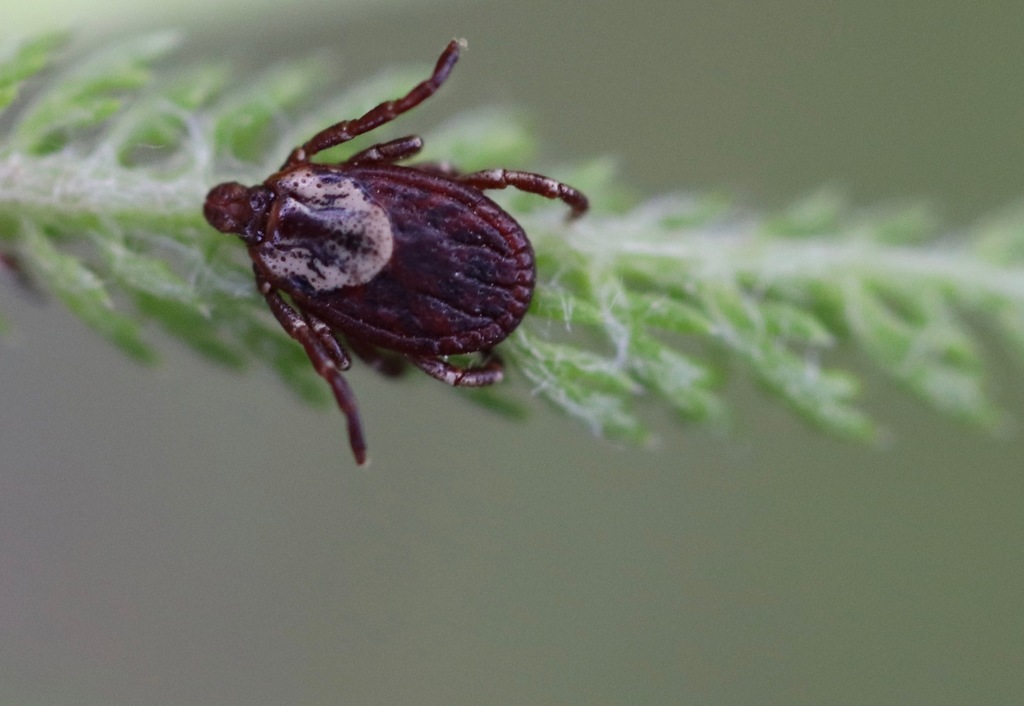
Regional Distribution & Vector Identification

Blacklegged Tick (Ixodes scapularis) showing reddish body and dark scutum — research-grade photo via iNaturalist
Ixodes scapularis · Research-grade · iNaturalist obs. #6211699
Blacklegged Tick
Ixodes scapularis
Regions
Northeast, Upper Midwest, Southeast
Visual ID
Reddish-brown body with a distinct, solid dark-brown oval scutum (shield); dark, blackish legs
Pathogens
Borrelia burgdorferi Anaplasma phagocytophilum Babesia microti Powassan virus
Western Blacklegged Tick (Ixodes pacificus) showing reddish body, dark scutum and prominent mouthparts — research-grade photo via iNaturalist
Ixodes pacificus · Research-grade · iNaturalist obs. #5597871
Western Blacklegged Tick
Ixodes pacificus
Regions
Pacific Coast (California, Oregon, Washington)
Visual ID
Highly similar to I. scapularis; oval scutum with long, prominent mouthparts
Pathogens
Borrelia burgdorferi Anaplasma phagocytophilum
Lone Star Tick (Amblyomma americanum) adult female with distinctive white star spot on scutum — research-grade photo via iNaturalist
Amblyomma americanum · Research-grade · iNaturalist obs. #227923559
Lone Star Tick
Amblyomma americanum
Regions
Southeast, Mid-Atlantic, Midwest, Northeast (expanding)
Visual ID
Rounder body; adult females have a single highly visible white spot ("star") on the center of the scutum
Pathogens
Ehrlichia chaffeensis Ehrlichia ewingii Francisella tularensis Heartland virus Bourbon virus Alpha-gal carbohydrate
American Dog Tick (Dermacentor variabilis) showing ornate white/grey dorsal scutum markings — research-grade photo via iNaturalist
Dermacentor variabilis · Research-grade · iNaturalist obs. #27055599
American Dog Tick
Dermacentor variabilis
Regions
East of the Rocky Mountains, portions of the Pacific Coast
Visual ID
Ornate, wider body with white/silver-grey ornamental striping patterns across the scutum
Pathogens
Rickettsia rickettsii Francisella tularensis
Rocky Mountain Wood Tick (Dermacentor andersoni) — research-grade photo via iNaturalist
Dermacentor andersoni · Research-grade · iNaturalist obs. #28270366
Rocky Mountain Wood Tick
Dermacentor andersoni
Regions
Intermountain West, Rocky Mountain states at high elevations
Visual ID
Similar to American Dog Tick but with brighter cream-colored ornamentation on the dorsal shield
Pathogens
Rickettsia rickettsii Francisella tularensis Colorado tick fever virus
Section 2

Clinical Classification of Pathogens

Class Pathogen(s) Primary Vector(s)
Bacteria — Spirochete Borrelia burgdorferi, Borrelia mayonii Ixodes scapularis, Ixodes pacificus
Bacteria — Obligate Intracellular Anaplasma phagocytophilum, Ehrlichia chaffeensis, Rickettsia rickettsii Ixodes spp., Amblyomma americanum, Dermacentor spp.
Bacteria — Gram-Negative Coccobacillus Francisella tularensis Dermacentor spp., Amblyomma americanum
Protozoan Parasite Babesia microti Ixodes scapularis
Virus Powassan virus, Heartland virus, Bourbon virus, Colorado Tick Fever virus Ixodes spp., Amblyomma americanum, Dermacentor spp.
Section 3

Clinical Data, Presentation & Diagnostics

Lyme Disease

Borrelia burgdorferi — Vector: Ixodes scapularis / Ixodes pacificus
Incubation
3–30 days post-bite
Early Presentation
Erythema migrans "bull's-eye" rash (≥5 cm expanding annular rash), systemic flu-like malaise, fatigue, arthralgia, fever
Late / Disseminated
Neurological abnormalities (facial palsy, radiculopathy, meningitis), migratory large-joint arthritis (especially knee), Lyme carditis (AV block — may require temporary pacing)
Diagnostics
Two-tier serological testing: ELISA screening followed by confirmatory Western Blot. Early EM is diagnosed and treated clinically — serology is frequently negative in the first 2–6 weeks.

Rocky Mountain Spotted Fever (RMSF)

Rickettsia rickettsii — Vector: Dermacentor variabilis / Dermacentor andersoni
Incubation
2–14 days post-bite
Early Presentation
High sudden fever, severe retro-orbital headache, abdominal pain, nausea, myalgia
Late / Severe
Maculopapular rash appearing 2–5 days post-fever onset — begins on wrists/ankles, spreads centrally; often involves palms and soles. Progresses to petechiae, DIC, multi-organ failure if untreated.
Diagnostics
PCR of skin biopsy or whole blood during acute febrile phase; Indirect Immunofluorescence Assay (IFA) using paired acute and convalescent serum titers (≥4-fold rise is confirmatory)
⚠ Do NOT delay treatment pending confirmatory results. RMSF mortality is significantly higher when doxycycline is started after day 5 of illness. Treat empirically on clinical suspicion.

Anaplasmosis & Ehrlichiosis

Anaplasma phagocytophilum / Ehrlichia chaffeensis — Vectors: Ixodes spp. / Amblyomma americanum
Incubation
5–14 days post-bite
Presentation
Fever, chills, severe headaches, myalgia, leukopenia, thrombocytopenia, elevated hepatic transaminases. Rash is rare in anaplasmosis but occurs occasionally in pediatric ehrlichiosis.
Diagnostics
Whole blood PCR during first week of illness; microscopic visualization of morulae (bacterial inclusions) inside leukocytes — neutrophils for anaplasmosis, monocytes for ehrlichiosis

Babesiosis

Babesia microti — Vector: Ixodes scapularis
Incubation
1 to several weeks post-bite (range variable)
Presentation
Hemolytic anemia, jaundice, dark urine, splenomegaly, profound fatigue, fever, sweats. Can be life-threatening in asplenic, immunocompromised, or elderly patients.
Diagnostics
Thin and thick peripheral blood smears stained with Giemsa or Wright stain — look for intraerythrocytic parasites and characteristic "Maltese cross" (tetrad) ring forms. Whole blood PCR is confirmatory and more sensitive in low-parasitemia cases.

Powassan Virus Encephalitis

Powassan virus (Flavivirus) — Vector: Ixodes scapularis / Ixodes cookei
Transmission Speed
15–30 minutes from tick attachment (critically faster than Lyme — no 36-hour safe window)
Presentation
Sudden high fever, altered mental status, seizures, focal neurological deficits, cranial nerve palsies. May range from asymptomatic to severe neuroinvasive meningitis or encephalitis.
Prognosis
~10% mortality in neuroinvasive cases; up to 50% of survivors suffer long-term irreversible neurological sequelae
Management
Strictly supportive care: manage intracranial pressure, IV hydration, seizure control, airway protection. No antiviral therapy is approved.
Section 4

Pharmacological Treatment Guide

First-Line: Doxycycline

Indications: Lyme disease, RMSF, Anaplasmosis, Ehrlichiosis

Adult dose: 100 mg PO or IV twice daily
Duration — Lyme (early localized): 10–14 days
Duration — Lyme (disseminated/arthritis): 14–21 days (oral); neurological Lyme: 14–28 days IV ceftriaxone
Duration — RMSF, Anaplasmosis, Ehrlichiosis: Minimum 7 days, continue at least 3 days after defervescence

Pediatric dose: 2.2 mg/kg per dose twice daily (max 100 mg per dose)
Doxycycline is first-line for RMSF in children of all ages — short courses (≤21 days) do not cause clinically significant dental staining.

Lyme Disease Alternatives (Pregnancy or Tetracycline Allergy)

Amoxicillin: 500 mg PO three times daily × 14–21 days
Cefuroxime axetil: 500 mg PO twice daily × 14–21 days
Note: Neither amoxicillin nor cefuroxime is effective for RMSF, Anaplasmosis, or Ehrlichiosis.

Babesiosis

Standard (mild–moderate):
Atovaquone 750 mg PO BID + Azithromycin 500 mg PO day 1, then 250 mg daily × 7–10 days

Severe / Immunocompromised:
Clindamycin 600 mg IV TID (or 300–600 mg PO TID) + Quinine 650 mg PO TID × 7–10 days
Consider exchange transfusion for parasitemia >10%, severe hemolysis, or cardiopulmonary compromise.

Viral Tick-Borne Diseases (Powassan, Heartland, Bourbon, Colorado Tick Fever)

Strictly supportive care only. No approved antiviral therapy. Antibiotics are ineffective. Manage intracranial pressure, maintain airway, IV hydration, seizure prophylaxis as indicated.
Section 5

Alpha-gal Syndrome (AGS) Clinical Protocol

Trigger: Galactose-alpha-1,3-galactose (alpha-gal) carbohydrate transferred via Lone Star Tick (Amblyomma americanum) saliva, sensitizing the host's immune system to mammalian-derived alpha-gal epitopes.

Diagnostic Criteria

  • Delayed reaction window: Symptoms appear 3–8 hours after ingestion of mammalian meat or products (due to metabolic clearance timeline of lipid-bound carbohydrates — distinguishes AGS from immediate IgE food allergy)
  • Symptom profile: Urticaria, angioedema, severe abdominal cramping, vomiting, diarrhea, or full systemic anaphylaxis
  • Lab: Serum Alpha-gal specific IgE (sIgE) blood test — value >0.1 IU/mL positive when aligned with clinical history
  • Skin prick limitation: Traditional skin-prick tests with raw meats frequently yield false negatives — serum sIgE testing is the preferred diagnostic modality

Long-Term Management

  • Primary avoidance: Strict elimination of mammalian meat (beef, pork, lamb, venison, bison). Poultry and seafood are safe.
  • Sensitivity-dependent secondary avoidance: Dairy products, gelatin-containing foods (marshmallows, gummies, gelcaps/medication capsules), lard — based on individual sIgE titer and reaction history
  • Emergency preparedness: Always prescribe and instruct on epinephrine auto-injector use (EpiPen or equivalent)
  • Prognosis: Subsequent Lone Star tick bites will spike sIgE levels and worsen sensitivity. Strict long-term avoidance of further bites can allow titers to naturally decline over several years.
Section 6

Emerging Tick-Borne Viruses

Powassan Virus (Flavivirus)

Vectors: Ixodes scapularis and Ixodes cookei (Groundhog tick)
Transmission
15–30 minutes from initial tick attachment — no extended attachment time required for transmission
Presentation
Ranges from asymptomatic to severe neuroinvasive disease (meningitis or encephalitis): sudden high fever, altered mental status, seizures, focal neurological deficits, cranial nerve palsies
Prognosis & Management
~10% mortality in neuroinvasive cases; up to 50% of survivors with long-term irreversible neurological sequelae. Management: strictly supportive care — no approved antiviral therapy.

Heartland & Bourbon Viruses

Vector: Primarily Amblyomma americanum (Lone Star tick)
Presentation
Highly mimics anaplasmosis/ehrlichiosis: fever, profound fatigue, anorexia, nausea, leukopenia, thrombocytopenia
Key Distinguishing Feature
Patients fail to respond to empiric doxycycline therapy — treatment failure in a suspected anaplasmosis/ehrlichiosis case should prompt viral workup
Management
Supportive care only. Notify state health department — both are notifiable conditions.
Section 7

Critical Diagnostic Pitfalls

Pitfall 1

Seronegative Window — Lyme Disease

Standard two-tier serology (ELISA + Western Blot) takes 2–6 weeks post-bite for IgM and IgG to reach detectable levels. Early localized Lyme with an erythema migrans rash must be diagnosed and treated clinically — waiting for laboratory confirmation yields high false-negative rates and delays treatment during the most treatable window.

Pitfall 2

Antibiotic Interruption of Seroconversion

Initiating doxycycline early can blunt the immune response, causing patients to remain seronegative on subsequent Western Blot testing even when infection was present. A negative follow-up serology in a clinically treated patient is not reliable evidence of absence of infection.

Pitfall 3

Tick Testing Limitations

The CDC explicitly discourages using commercial tick PCR testing to guide clinical decisions. Pathogen presence in the tick does not guarantee transmission occurred; a negative test does not rule out infection from concurrent unknown bites. Treatment must be guided by patient symptomatology and epidemiological exposure history — not tick test results.

Pitfall 4

Rickettsial Cross-Reactivity on IFA Testing

IFA testing for RMSF (Rickettsia rickettsii) exhibits significant cross-reactivity with other spotted-fever group rickettsiae (e.g., Rickettsia parkeri, R. amblyommatis), which cause less severe illness. Definitive species-level diagnosis may be challenging serologically. Treatment protocols and urgency remain identical regardless of the specific rickettsial species.

Section 8

Post-Bite Action Plan

Step 1 — Mechanical Removal

Use fine-tipped tweezers. Grasp the tick's mouthparts as close to the skin surface as possible. Pull upward with steady, even pressure — do not jerk or twist. Do not squeeze the tick's body. Do not apply heat, petroleum jelly, nail polish, or any chemical — these methods are ineffective and may increase pathogen transmission risk.

Step 2 — Wound Disinfection

Clean the bite site immediately with soap and water, rubbing alcohol (isopropyl 70%+), or iodine scrub. Dispose of the tick by placing in sealed container, submersing in alcohol, or flushing down the toilet.

Lyme Disease Prophylaxis — Single-Dose Doxycycline 200 mg PO

Prophylaxis is indicated when ALL of the following criteria are met:

  1. Tick reliably identified as adult or nymphal Ixodes scapularis (Blacklegged tick)
  2. Estimated attachment duration ≥ 36 hours (based on degree of engorgement or credible exposure timeline)
  3. Local ecological infection rate of B. burgdorferi in host ticks is ≥ 20% (applies to highly endemic Northeast and Upper Midwest regions)
  4. Prophylaxis can be started within 72 hours of tick removal
  5. No contraindications to doxycycline (pregnancy, age <8 years)
Single-dose prophylaxis is NOT indicated for Western Blacklegged tick (I. pacificus), Lone Star tick, or Dermacentor species bites — insufficient evidence supports efficacy in those exposures.
Section 9

State-by-State Public Health Reporting Registry

All listed tick-borne diseases are nationally notifiable conditions. Report confirmed and probable cases to your state health department within the required timeframe.
AL — Alabama
Department of Public Health (Epidemiology)
1-800-338-8374
AR — Arkansas
Department of Health (Zoonotic Disease)
1-501-280-4186
CA — California
Department of Public Health (Vector-Borne)
1-916-552-9730
CT — Connecticut
Agricultural Experiment Station (CAES) Tick Lab
1-203-974-8500
DE — Delaware
Division of Public Health
1-888-295-5156
FL — Florida
Department of Health (Bureau of Epidemiology)
1-850-245-4401
GA — Georgia
Department of Public Health (Epidemiology)
1-404-657-2700
IA — Iowa
Department of Health and Human Services
1-800-362-2736
IL — Illinois
Department of Public Health
1-217-782-2016
IN — Indiana
Department of Health (Zoonotic)
1-317-233-7125
KS — Kansas
Department of Health and Environment
1-877-427-7317
KY — Kentucky
Department for Public Health
1-502-564-3261
LA — Louisiana
Department of Health
1-800-256-2748
MA — Massachusetts
Department of Public Health
1-617-983-6800
MD — Maryland
Department of Health (Infectious Disease)
1-410-767-6700
ME — Maine
Center for Disease Control and Prevention
1-800-821-5821
MI — Michigan
Department of Health and Human Services
1-515-242-5935
MN — Minnesota
Department of Health (Vector-Borne)
1-651-201-5414
MO — Missouri
Department of Health and Senior Services
1-573-751-6113
MS — Mississippi
State Department of Health
1-866-458-4948
NC — North Carolina
Division of Public Health
1-919-733-3419
NE — Nebraska
Department of Health and Human Services
1-402-471-2937
NH — New Hampshire
Department of Health and Human Services
1-603-271-4496
NJ — New Jersey
Department of Health
1-609-826-5964
NY — New York
State Department of Health Vector-Borne Diseases
1-518-474-4568
OH — Ohio
Department of Health (Zoonotic)
1-614-752-1029
OK — Oklahoma
State Department of Health
1-405-426-8710
PA — Pennsylvania
Department of Health Bureau of Epidemiology
1-877-724-3258
RI — Rhode Island
Department of Health
1-401-222-2577
SC — South Carolina
Dept of Public Health
1-800-922-0204
TN — Tennessee
Department of Health
1-615-741-7247
TX — Texas
Department of State Health Services (Zoonosis)
1-512-776-7111
VA — Virginia
Department of Health
1-800-533-4148
VT — Vermont
Department of Health
1-802-863-7240
WI — Wisconsin
Department of Health Services
1-608-266-1120
WV — West Virginia
Department of Human Services
1-304-558-5358